
Supplementary Files
Keywords
meniscus
swine model
biomechanics
scaffolds
How to Cite
Abstract
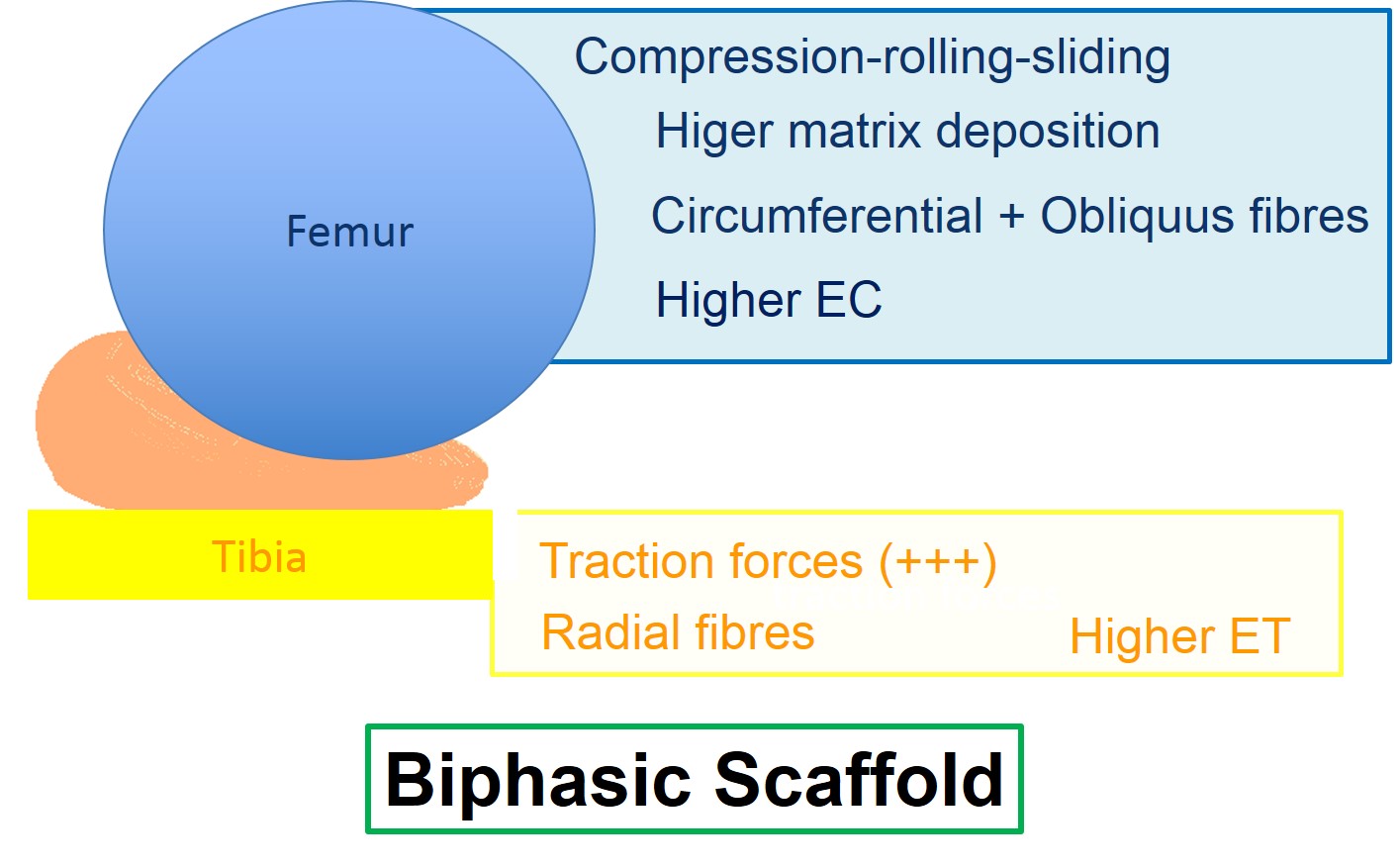
Menisci are wedge-like structures interposed, in the knee joint, between the femoral and the tibial articular heads (Kohn et al. 1995; Greis et al. 2002). Improving the articular surface, the cellular nutrition and the articular lubrication, they are essential structures for the prevention of gonarthrosis (Proctor et al.1989; Makris et al. 2011). This study is focused upon the relationship between the contact forces at the femoral and tibial surfaces and the corresponding structure of these meniscal surfaces. For this purpose, 20 adult (~9 months old) female pigs (Landrace x Large white, average weight 75–90 kg; n=80 meniscal samples) were obtained from a local slaughterhouse and dissected to isolate the menisci. Swine meniscal samples were evaluated from morphological (Safranin-O, Sirius Red and collagen type I and II) (Di Giancamillo et al. 2014), biochemical (DNA and glycosaminoglycans, or GAGs, contents) and biomechanical (compression and traction tests) points of view at the level of femoral and tibial meniscal surfaces. Results revealed a characterization of the meniscus which is biomechanical-dependent. The femoral surface, morphologically characterized by the interposition of radial and oblique fibers and biomechanically by the femoral condyles compression, sliding and rolling forces, shows a higher compressive modulus (p<0.05) and a greater amount of cells and GAGs deposition (p<0.01 for each analysis). On the other hand, results from traction test revealed a higher tensile modulus (p<0.05) in the tibial surface, characterized by a circumferential arrangement of the fibers and a poorer GAGs deposition and cellular distribution (p<0.01). Results (summarized in the figure 1) from this work suggest that a biphasic “femoral-to-tibial” scaffold that mimic the different behavior and composition of the two meniscal surfaces could be useful in the light of meniscal replacement.
References
Di Giancamillo, A., Deponti, D., Addis, A., Domeneghini, C. and Peretti, G.M., 2014. Meniscus maturation in the swine model: changes occurring along with anterior to posterior and medial to lateral aspect during growth. Journal of Cellular and Molecular Medicine. Journal of Cellular and Molecular Medicine. 10: 1964-1974.
Greis, P.E., Bardana, D.D., Holmstrom, M.C., and Burks, R.T. 2002. Meniscal injury: I. Basic science and evaluation. Journal of American Academy of Orthopaedic Surgery. 10(3):168-76.
Kohn, D., and Moreno, B., 1995. Meniscus insertion anatomy as a basis for meniscus replacement: a morphological cadaveric study. Arthroscopy. 11(1):96-103.
Makris, E.A., Hadidi, P., and Athanasiou, K.A. 2011. The knee meniscus: structure-function, pathophysiology, current repair techniques, and prospects for regeneration. Biomaterial. 32(30): 7411–7431.
Proctor, C.S., Schmidt, M.B., Whipple, R.R., Kelly, M.A., and Mow, V.C. 1989. Material properties of the normal medial bovine meniscus. Journal of Orthopaedic Research. 7(6):771-82.
This work is licensed under a CC BY-SA 4.0 international